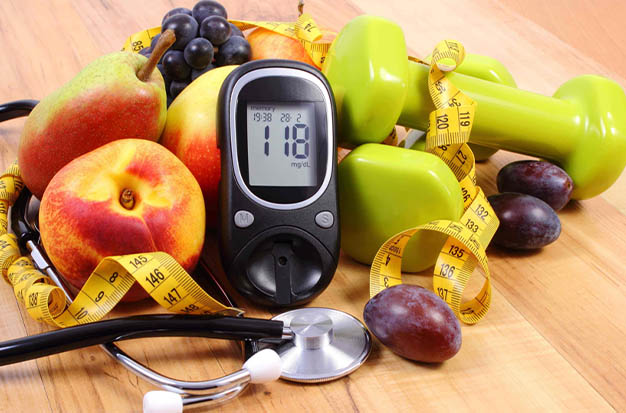
How Can You Tell Where & When Sugar Enters The Blood
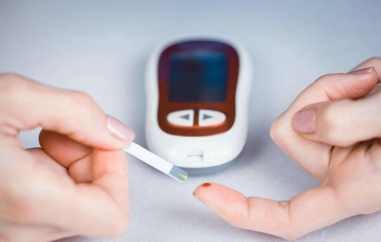
Typically, the ventral side of the finger is the area where the blood glucose meter measures blood sugar. The blood collection component needs to be cleaned prior to the test. You can wash your hands in warm water with soap after collecting blood, and then dry them with cotton balls or clean napkins. After cleaning, the arm where the blood collection part is located should hang naturally for a short while. Then, the blood collection part should be massaged, and a sufficient number of blood samples should be obtained using an appropriate blood collection device.
Avoid squeezing the blood sampling site because doing so will dilute the blood sample and affect the outcome of the blood glucose test. It is advised to draw enough blood at one time for the test. The blood glucose test paper and blood glucose meter should not be touched or moved while the test is being performed.
What Is Glucose?
Glucose, which serves as the body’s primary source of energy, is found throughout the entire body. Blood glucose is currently the most widely recognized form of human glucose, but cerebrospinal fluid, tissue fluid, and tears are also abundant sources of this compound! The body’s various tissues and cells directly use this non-blood glucose, which is closely related to blood glucose.
Since humans have evolved over millions of years, their ability to use glucose as an energy source has allowed them to live a life that is fundamentally a process of converting matter into energy. The burning of glucose by human body cells is what has given rise to the activity of human life.
The conversion of dietary carbohydrates, proteins, and fats into glucose results in a stable level of glucose in the body. It can leave the body in two ways: it can be eliminated through urine, or it can be converted by cells into carbon dioxide and water (a process that necessitates the involvement and activation of insulin).
We previously mentioned blood glucose, which is the glucose present in the blood of a living organism. Various tissue cells receive the glucose through the medium of blood. The majority of the cells, however, cannot directly contact the blood, which means they use glucose last. Only the glucose in the tissue fluid, also known as tissue fluid sugar, is what they come into direct contact with.
The relationship between blood glucose and tissue liquid sugar is very straightforward and clear because blood glucose can enter the tissue fluid through easy infiltration and diffusion to become tissue liquid sugar.
Because the two have achieved osmotic balance, it is almost possible to say that tissue liquid sugar is equivalent to blood sugar in the steady state of human fasting.
Tissue liquid sugar will rise more slowly than blood sugar when blood sugar increases rapidly as a result of human consumption because tissue liquid sugar is a downstream effect. But both have a consistent upward trend.
The body will quickly enter the cells for cellular oxidation and utilization of the tissue liquid sugar when it senses a sharp rise in blood sugar levels and secretes insulin to encourage cell utilization of glucose. The tissue liquid sugar will be lower than the blood sugar at this time because the blood sugar and tissue liquid do not enter synchronously. After the tissue liquid is given, however, the blood sugar will also be lower.
Blood must be drawn through broken blood vessels in order to measure blood sugar; however, collecting tissue fluid to measure tissue fluid sugar requires less trauma because tissue fluid is distributed beneath the skin, where blood vessels are distributed much more shallowly.
The change in blood sugar can be detected through the monitoring of tissue liquid sugar due to the close association between blood sugar and it, as well as the consistency of their respective change trends. The time difference between blood sugar and blood sugar when the blood sugar changes quickly and the reasons for the difference, however, must be taken into consideration when interpreting tissue liquid sugar.
Blood Glucose Source
(1) consuming and absorbing carbohydrates: starch and glycogen in food are decomposed by amylase to release glucose and then absorbed by the digestive tract, which is the main source of blood sugar.
(2) Glycogen breakdown: after short-term starvation, glycogen stored in the liver and muscle is decomposed into glucose and enters the blood, which is glycogen decomposition.
(3) Gluconeogenesis: In the liver, non-sugar substances like amino acids and glycerol use gluconeogenesis to produce glucose in response to prolonged starvation.
How Does The Body Use Blood Sugar?
(1) Oxidative decomposition glucose generates The main way that blood glucose enters the body is through the production of ATP in tissue cells through aerobic oxidation and anaerobic fermentation.
(2) After a meal, tissues like the liver and muscle synthesize glucose into synthetic glycogen for storage.
(3) Changing to non-sugar substances: conversion to glycerol and fatty acids to synthesize fats; used to create proteins by converting to amino acids.
(4) It is changed into various other sugars and sugar derivatives, including ribose, deoxyribose, amino polysaccharide, etc.
(5) whenever the blood glucose level exceeds the renal threshold (8.9-10mmol / L, 160–180 mg/dl), can be excreted in part in the urine.
Where Is The Sugar Screen’s Blood-drawing Component?
The most conventional and efficient method of doing so is with a glucose meter You prick your finger with a needle and wash your arms to remove germs.
The original purpose of sugar screening was to examine the likelihood of fetal malformation through blood sampling and a serum separation test. Therefore, the blood sampling component is important to many expectant mothers. In actuality, the blood sample portion is identical to other tests. Only the elbow vein must be chosen in order to take blood. Typically, 4ml of blood is drawn.
Blood is drawn to check the sugar screen. Some claim they are unsure of how much blood to draw or from where to draw it for this test. We will therefore examine the portion of the blood sample to be drawn and the sugar screen today. I’m hoping that everyone can grasp the sugar screen’s fundamental concepts.
Let’s start by examining the area where the test draws blood. For this test, the vein at your elbow, which is located in the middle of your arm, is typically where blood will be drawn from. There is no set rule regarding which arm should be used. Therefore, you can choose the arm from which you think it will be most convenient to take a blood sample from the elbow vein.
So, how much blood is required for a sugar screening? The amount of blood required for this test is only about 4ml, so this is a reasonable amount to draw.
What then is the mechanism by which sugar screening eliminates fetal malformations? In order to achieve the goal of screening, the appropriate units will draw serum from your blood, analyze the concentration of AFP and hCG present in the serum, and then combine these values with the anticipated delivery date, gestational week, age, weight, and other characteristics of the pregnant mother to determine how high the risk factor is.
We must focus especially on a specific aspect when examining the risk coefficient of sugar screening. It is correct to assume that the risk increases as the number decreases. Its accuracy, though, isn’t particularly good. The accuracy rate is less than 80% under typical conditions. In other words, the results of this inspection were either critical or high risk, necessitating additional inspection and judgment.
Where did the test’s blood draw take place? The solution was already discussed. You must be aware of some pertinent information about this exam in advance if you plan to take it. Making the necessary preparations requires a thorough understanding of the pertinent information. You can only use this examination more effectively in this way if you want to get the best results possible in terms of eliminating deformities.


Average Rating